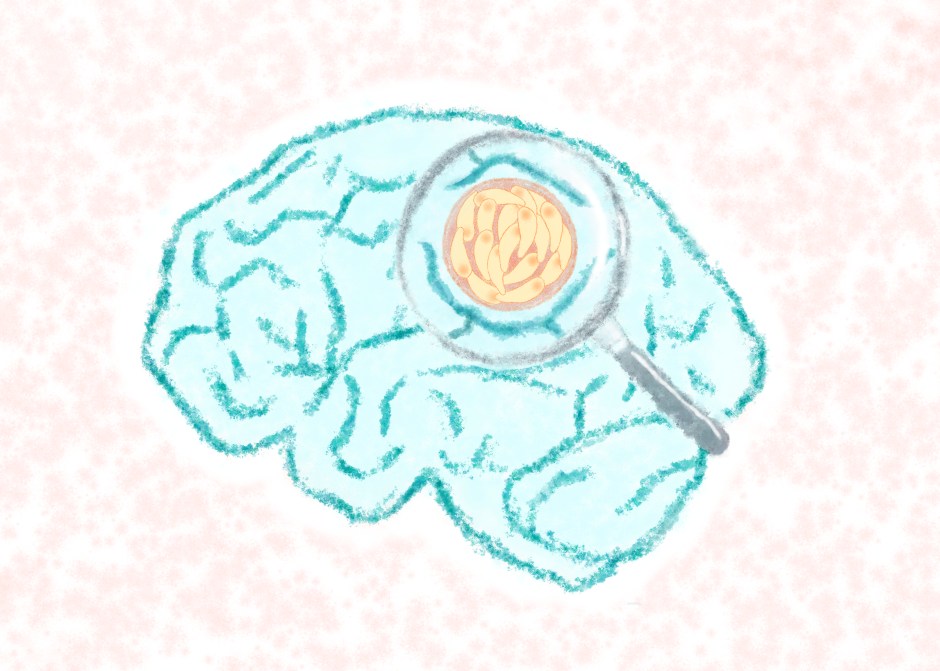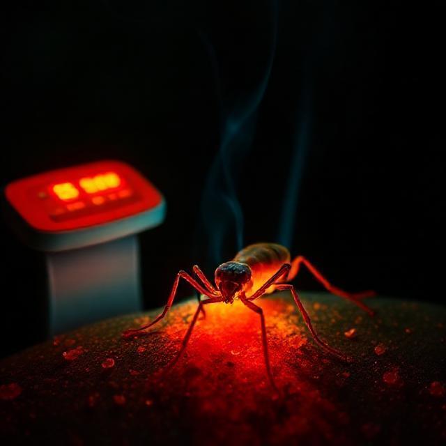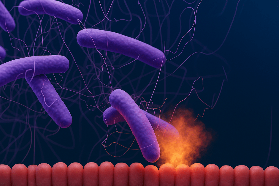
Breaking down the microbiology world one bite at a time
Martyrs of infection
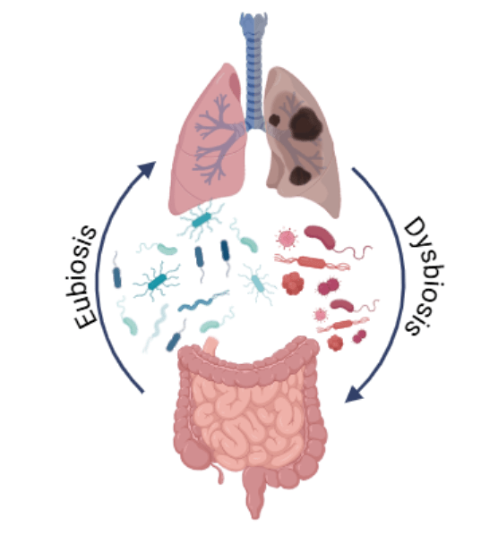
Intestine’s skin: a barrier against pathogens
Think about a wound you got from a knife while cutting vegetables or maybe a burn when you touched a searing hot steel saucepan. How painful it was! Now, remember how a thin layer of skin develops on the wound over time, reducing the pain to almost nothing. Just like the outer skin, all mammals have an inner skin lining for all internal organs like lungs, liver, pancreas, stomach and intestine. This layer, called the epithelium, protects mammals against internal infection and damage through a variety of mechanisms.
A lot of current research is focused on intestinal epithelium because the cells of this layer work in mysterious ways. Pathogens target one cell at a time by invading them through needle-like structures, making their way to other internal organs through the network of cells and blood. But, wait! It is not so easy to disrupt the harmony of the mammalian body. Intestinal epithelium serves as the first barrier to this disruption. When one cell is invaded by the pathogen, it communicates with the neighboring through cell-signaling pathways, forming a network almost like a WiFi hotspot internet connection. Amongst the neighboring cells are also the ones forming muscles surrounding the intestine. Until now researchers thought that the muscles help in contracting the intestinal wall when a pathogen attacks, but they did not know that the cells of the intestinal epithelium can also defend against the attack. Recently, researchers from Uppsala University have found that infected epithelial cells can withstand infection on their own and signal the surrounding epithelium cells to not get affected during this process. These researchers conducted an experiment to back their findings, where they saw that the epithelium layer undergoes contraction at the site of infection and sheds the affected cells.
Cultivating skin in a petri dish!
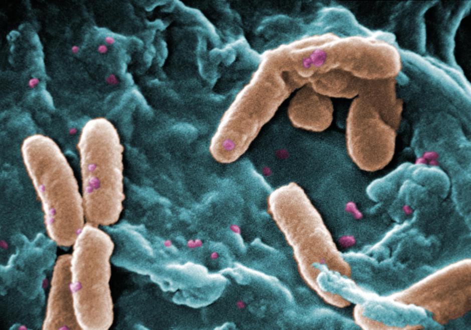
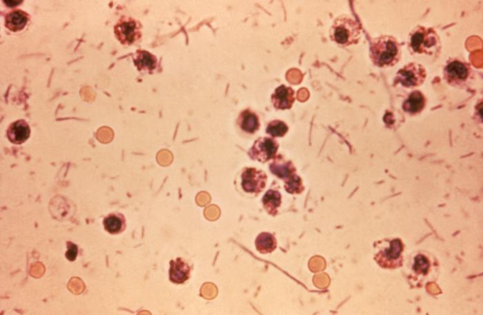
Yes! All those sci-fi movies might be true after all! Scientists can cultivate live tissues on a petri dish, with a varying degree of density. In this particular case, they created a single layer of epithelial cells derived from the intestines of mice. These cells transform into sheet-like tissues without any openings on a plastic surface over time. These sheets are called enteroids. Once the cells form enteroids, scientists infect them with Salmonella Typhimurium (STm) to study the mechanism of invasion and subsequent epithelial response. STm is a pathogen known for causing typhoid-like infection and uses a needle-like structure to invade the intestinal epithelial lining. If STm is devoid of the genes that code for proteins of the needle, it cannot induce any epithelial immune response.
How does the mammalian intestine protect itself against harmful bacteria?
During the pathogen attack on the epithelium, the affected cells can lyse and harm the integrity of the intestinal wall further by disintegrating the surrounding cells. Think of it like a wooden shed damaged by rain on the outside. It is not long before the damage quickly spreads to the rest of the wood including the underside of the shed and the entire structure collapses. But the intestinal epithelial cells are smart! During infection, cells that get invaded are expelled from the epithelial layer and die (Figure 1). In this process, they act as saviors for the cells surrounding them. How so?

Researchers prepared enteroids from mice intestines which lacked crucial genes (Casp1/11 and GsdmD) that induced contractions in the epithelium layer. They then infected these enteroids with Salmonella Typhimurium and compared it to the infected enteroids from mice with all genes intact (Figure 2). Surprisingly, researchers found that the cells from deficient enteroids still died in the absence of contractions. Yet, these enteroids occupied lesser space than the enteroids with all genes intact. This proves that in the absence of contractions, the epithelial layer integrity is compromised, much like the wooden shed. But, in the presence of contractions, cells tend to form a densely packed area at the site of invasion. This process leads to literal squeezing of the dead infected cells and some live cells from the layer. What happens to the gap created, you will ask now? That’s where the densely packed structure comes in. The gap is filled by the neighboring cells because there were too many of them at the infection site, to begin with. As for what happens to the pathogens, that is a story for another time!

Like any other experiment in mice, there is always a question: Does this also happen in humans? In this case, researchers developed human intestinal enteroids derived from tissues extracted during bariatric surgery and infected them STm. They found that the effect observed in mouse-derived enteroids occurred at a lesser intensity in human-derived enteroids.
Sacrifice of intestinal epithelial soldiers
Mammalian body is made of a number of cells, where each cell has a purpose, just like a jigsaw puzzle. These cells work in mysterious ways. In this study, researchers have tried to unfold one of these mysteries at a microscopic level. The epithelium cells work in a pack. To maintain the integrity of the whole pack, some cells sacrifice themselves, becoming martyrs in the battle of infection. In the long run, this sacrifice prevents further disintegration of the surrounding tissues and subsequent systemic infection.
Other references:
Eisenhoffer GT, Loftus PD, Yoshigi M, Otsuna H, Chien CB, Morcos PA, Rosenblatt J. Crowding induces live cell extrusion to maintain homeostatic cell numbers in epithelia. Nature. 2012 Apr 15;484(7395):546-9. https://doi.org/10.1038/nature10999
Featured image: Author’s design