
Breaking down the microbiology world one bite at a time
Mosquitoes, Midguts and Malady
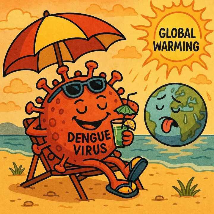
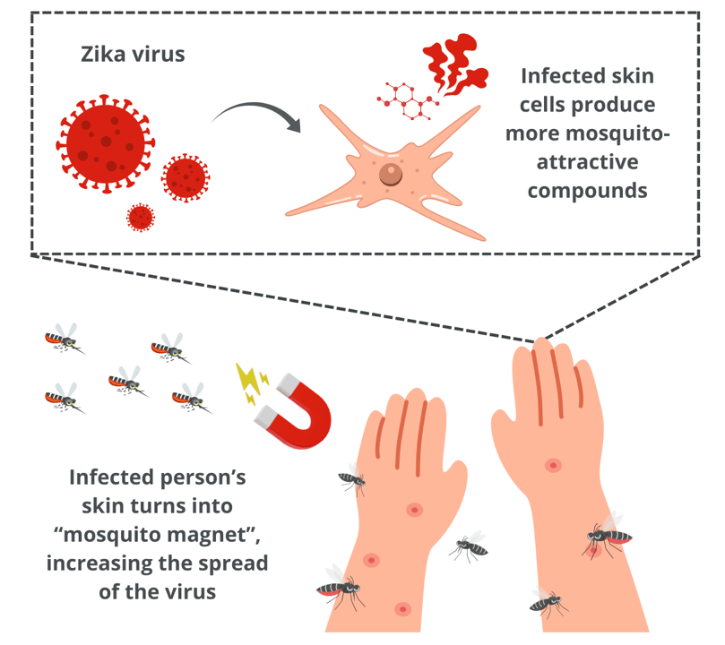
It’s difficult to name a more infamous pest than the creature known as the mosquito. Not only do these flying insects ruin summer picnics, but they also transmit numerous diseases. A highly efficient species is the Aedes aegypti mosquito that can transmit numerous diseases including Dengue virus, Yellow fever virus, Chikungunya virus and Zika virus.
One integral component of disease outbreak modeling and vector control programs is understanding the feeding behaviors of the vectors that transmit the disease among hosts. In the case of the mosquito, this means an understanding of what happens after a mosquito bites or takes a blood meal from an infected host and then feeds from a different host.
The Aedes aegypti mosquito itself has a relatively high feeding frequency, biting humans at a rate of 0.63 to 0.76 times per day. These blood meals may be considered partial or full depending on the feeding success of the mosquito which can be interrupted by host defenses such as swatting it away. If the mosquito is unable to take a complete meal, the stretch receptors in the mosquitoes’ midgut, and the degree to which the midgut is distended signals the mosquito to continue host seeking behaviors.
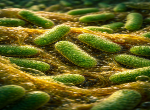
When feeding on a virally infected person, the blood the mosquito takes contains the virus particles and thus, disseminates into the mosquito’s midgut. On a subsequent blood meal, the virus can be transmitted to an uninfected host if the virus escapes through the midgut barrier which is primarily formed by a protein called type IV collagen.
These aspects of viral transmission dynamics are very much underexplored and the researchers in this article seek to understand the interplay between midgut collagen damage, blood meal size and viral dissemination and transmission.
To investigate midgut damage, scientists conducted a Collagen Hybridization Peptide (CHP) Assay. Collagen is a highly abundant protein used to strengthen and support connective tissue. Due to its abundance in the mosquito midgut, damage to the type IV collagen barrier means loss of integrity of the midgut and release of its components. In the CHP assay, midguts were dissected and prepped, then incubated with a protein that binds to denatured or broken-down collagen. This protein contains a fluorescent compound that can then be used to measure the levels of broken-down collagen using fluorescent microscopy.
When given a partial infectious (Dengue virus) blood meal, the mosquito’s midgut sees less expansion than those given a full blood meal. In addition, there are significant differences in the amount of collagen damage 24 hours post blood meal (hbpm) between the unfed controls, partially fed and fully fed groups.

Additionally, groups given only one single full blood meal had less midgut damage than those given a full initial meal and a secondary partial meal and those given 2 full meals. Regardless of secondary feeding state or volume, the infection prevalence among the groups was similar at 7 days post infection.
However, the presence of a secondary blood meal impacted the Dengue virus (DENV) dissemination; having a higher prevalence of DENV dissemination than without a secondary blood meal (no differences between partial vs full) at 7 days post infection.
As evidenced by these experiments, collagen IV damage in the mosquito midgut is directly influenced by blood meal size and subsequent midgut expansion. Furthermore, the acquisition of a secondary blood meal has an extensive effect on virus dissemination. This delicate interplay between volumes of blood meals and the need for subsequent feeds is a huge component in the spreading of vector-transmitted diseases. By highlighting this connection, we can better design vector control programs, outbreak models and even future laboratory experiments. One component that still needs exploration, however, is how the virus itself escapes the mosquito midgut and if damage or stress is even a necessary component for the escape to occur.
Link to the original post:
Increased blood meal size and feeding frequency compromise Aedes aegypti midgut integrity and enhance dengue virus disseminationJohnson RM, Cozens DW, Ferdous Z, Armstrong PM, Brackney DE (2023) Increased blood meal size and feeding frequency compromise Aedes aegypti midgut integrity and enhance dengue virus dissemination. PLOS Neglected Tropical Diseases 17(11): e0011703. https://doi.org/10.1371/journal.pntd.0011703
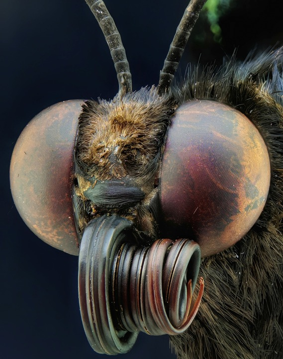
Featured image: Figure: Mosquito head. Image source: Photo by Agoenk Fatahillah.