
Breaking down the microbiology world one bite at a time
Museomics: A deep dive into museum specimens
During a recent visit to an ocean exhibit, I came across various specimens of deep-sea creatures, from preserved organisms in jars to skeletal structures and even live starfish. The exhibit also included interactive displays, one of which involved using a magnifying glass to observe plankton floating in collected seawater. Looking at these tiny creatures made me wonder what else could be discovered in such specimens at the microscopic level.
Similar to the ocean exhibit I visited, natural history museums across the globe host a wide range of preserved specimens and research collections from various environments, including biological samples collected decades ago. These specimens can harbour valuable genetic information that can be explored using modern sequencing technologies, such as metagenomics. Such studies fall under the field of museomics, which can be defined as the “study of genomic data obtained from ancient DNA and historic DNA specimens in museum collections.”
Museomics has been used to study preserved microbial DNA in museum specimens. In the past, it has helped researchers investigate the origins and spread of historical or ongoing diseases such as Lyme disease, leprosy, and malaria. DNA extracted from certain museum specimens can be used to identify specific pathogens that were present when the specimens were collected and preserved. Researchers have also used historical DNA to reconstruct the epidemiology of past diseases and investigate the evolution of pathogens such as Plasmodium, which causes malaria.
The use of museomics would significantly reduce the need for extensive environmental sampling, yet this method is not popular due to challenges related to foreign microbial contamination introduced during specimen collection, handling and preservation. Apart from studying pathogens, this method can also be adopted to identify uncultivable bacteria and host-specific microbial symbionts. Especially when it comes to small and rare marine animals that are difficult to collect from the vast oceans, this technique can come in handy.
Nudibranchs are shell-less molluscs with vibrantly coloured, soft bodies, commonly known as sea slugs. These organisms are studied for their unique chemical defense systems. Some nudibranchs steal stinging cells from their prey such as cnidarians, while other nudibranchs produce their own chemicals or acquire them from food. Around 3,000 species of nudibranchs are known so far, of which the microbiomes of only 23 species had been characterised previously.
Recently, the authors of this paper used museomics to investigate the microbiomes of nudibranchs using ethanol-preserved specimens archived for up to 30 years. Previous data revealed a symbiotic relationship between the nudibranch Doriopsilla fulva and the bacterium Candidatus Doriopsillibacter. Since then, Mária Džunková and colleagues have been on the lookout for similar symbionts in other nudibranch species using museum specimens.
They extracted microbial DNA from the mantle of 54 nudibranch specimens spanning 22 different species. Microbial diversity using the DNA samples was analysed using metagenomics. Microbial contamination was taken into consideration during sequencing analysis by examining the abundance, distribution, and identities of the detected bacteria. Of the 4,116 bacterial groups identified, 98.7% were considered likely to represent transient bacteria from seawater or microbes introduced during preservation and handling. The remaining 1.2% met the criteria for the core microbiome of the nudibranch specimens.
Out of the core microbiomes, the scientists identified dominant symbiotic associations for the two nudibranch genuses Polycera and Felimare. The associated bacterial lineages were the previously unknown uncultured bacterial lineages named Candidatus Polyceribacter and Candidatus Felimaribacter. Researchers further dived into the genomes of these symbionts and identified genes potentially involved in the production of compounds such as terpenes and polyketides. These compounds may play a role in the chemical defense of *Polycera* and *Felimare* nudibranchs, helping protect them against predators.
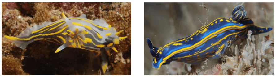
Combining their own observations with findings from previous studies, the researchers concluded that only some nudibranchs might rely on symbiotic relationships for defense and that these nudibranchs independently establish partnerships with distinct bacterial lineages. Some nudibranchs may instead obtain defensive compounds from their diet or produce them themselves.
Investigating marine invertebrates such as nudibranchs and their associated bacteria can provide us with valuable information about how host–microbe interactions occur within diverse marine ecosystems. In addition, these symbiotic bacteria might carry the genetic machinery needed to produce novel bioactive compounds with pharmaceutical potential. Relying solely on environmental samples could make such studies more challenging, as it may be difficult to collect a wide range of nudibranch species from different locations across the ocean. Therefore, this study also highlights how museum specimens and museomics can be used to investigate bacterial symbionts associated with rare or difficult-to-collect marine invertebrates.
Link to the original post: Porcel Sanchis, D., Pola, M., Engelberts, J.P. et al. Museomics reveals uncultured symbionts with biosynthetic potential in nudibranchs. Microbiome 14, 184 (2026). https://doi.org/10.1186/s40168-026-02456-z
Featured image: Vanessa Kunston – California Academy of Sciences