
Breaking down the microbiology world one bite at a time
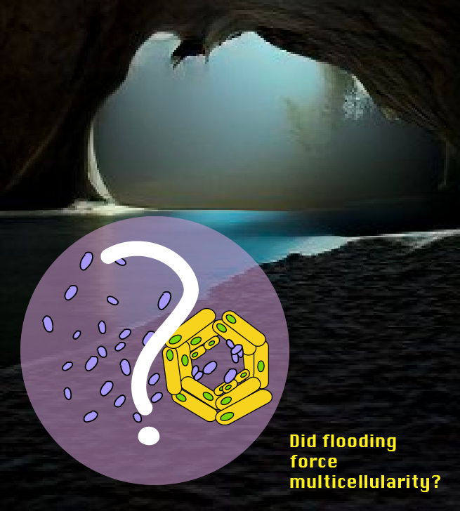
Did flooding force bacterial multicellularity?
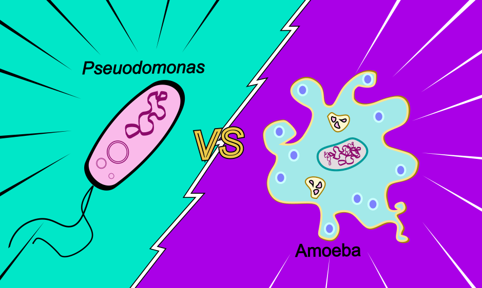
Bacteria, as we know them, are single celled organisms. This means that they can perform all their requisite functions within the context of a single cell. In contrast, humans are made up of about 37 trillion cells (that’s 37 followed by 12 zeros!). A single human cell would be lost on its own. How life went from one cell to multicellular is a hotly debated topic among scientists. Unfortunately, there are no neat answers to this huge question. But, fortunately for us, nature hides various clues to help us to solve this mystery.
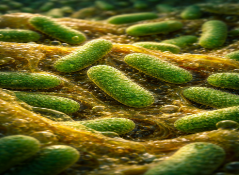
A limestone cave in the northern Kyushu Island of Japan was hiding one such clue: a fascinating new organism! A team of scientists in Japan sampled this organism from a site with unique ecological architecture: it was located slightly above the water surface of a river that flows underground. Periodically, when there is excess runoff post rainfall, the sampling site was submerged by the river. Realizing that this was a novel species, the scientists named it Jeongeupia sacculi sp. nov. HS-3.
New bacteria are being discovered all the time, so what’s so special about this cave microbe? Typically, bacteria grown in the lab on solid agar medium divide in a disordered fashion, generating opaque colonies. HS-3, however, seemed to produce transparent colonies with an iridescent hue! Upon looking closely, the scientists observed that this optical difference was a result of the unique organization of these colonies. They seemed to arrange closely in what resembled a liquid crystal.
Fascinated by this unique property, the team of scientists decided to observe the bacterial colony grow over the course of time. This continuous observation allowed them to distinguish different stages in the generation of HS-3’s colony. For the first 10 hours of growth, cells propagated as coccobacilli (the bacterial shape was somewhere between spherical and rod-shaped) (Figure 1A). Then, cells at the edge of the colony began to elongate and form a two-dimensional liquid crystal-like structure (Figure 1B). These colonies spread as filamentous cells in a single layer (Figure 1C). As it kept growing, bulges of cells became visible particularly at the edge of the colony, perhaps to relieve some of the pressure generated by internally dividing cells. After two days, the colonies stopped growing and seemed to make no changes for the next few days.

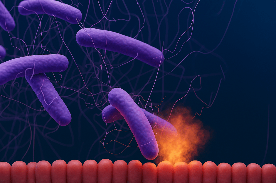
But HS-3 was far from done with its cycle. After resting for a couple of days, the cells in the center of the colony began proliferating and the colony started to become three-dimensional. The center of the colony turned from transparent to opaque indicating that something had changed about its internal structure. In contrast to the filamentous bacteria that made up the edges of the colony, the center consisted of coccobacilli, which were dividing disorderly (Figure 1D, Video 1).
Why were the internal cells so different from the rest of the cells in the colony? Turns out, the “heart” of these bacteria lay in their home a.k.a. in their environment. Since the site where these bacteria had been found was periodically submerged in water, the scientists did a simple, but elegant experiment. They took HS-3 colonies growing on agar plates and submerged them in water. Interestingly, those internal coccobacilli were immediately released into the water column (Figure 1E, Video 2), leaving behind the filamentous edges of the colony! Even after all the coccobacilli had been released, the filamentous cells at the edge stuck together tightly indicating that they were holding together to support the internal cells, allowing them to be released appropriately. Upon release, these coccobacilli may initiate HS-3’s cycle all over again, perhaps in a new uninhabited part of the cave. The differentiation of cell types in HS-3’s colony into filamentous and coccobacilli adapted to their ecological niche, providing a fascinating new way in which multicellularity can exist.
Although this work does not solve the problem of how multicellularity evolved, it helps us garner a new appreciation for how the environment can significantly influence organismal lifestyle.
Link to the original post: Kouhei Mizuno, Mais Maree, Toshihiko Nagamura, Akihiro Koga, Satoru Hirayama, Soichi Furukawa, Kenji Tanaka, Kazuya Morikawa (2022) Novel multicellular prokaryote discovered next to an underground stream eLife 11:e71920
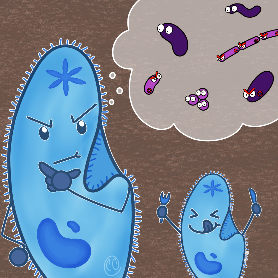
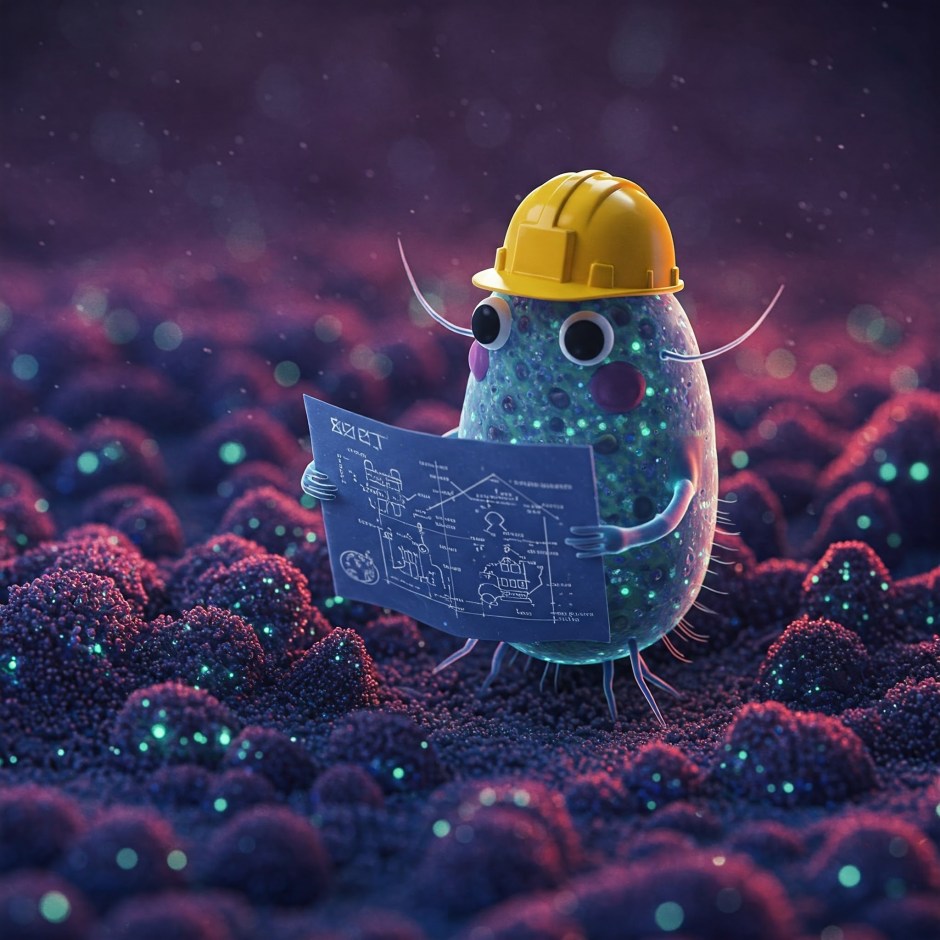
Featured image: Created by author using Adobe Illustrator and Craiyon