
Breaking down the microbiology world one bite at a time
Leishmaniasis Treatment Using Protein Destroyers
What is Leishmaniasis?
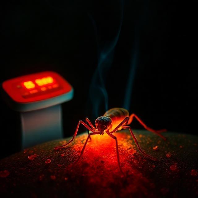
Leishmaniasis is an infection caused by the parasites from the Leishmania genus. These parasites are transmitted to humans through the bite of an infected female sandfly. Depending on the specific species of Leishmania, infection can manifest as either cutaneous, mucocutaneous or visceral leishmaniasis. Cutaneous leishmaniasis is the most common form, whereas visceral is the most severe, possibly leading to death if not treated.
What are Serine Proteases?
Proteases are enzymes (proteins that speed up reactions) that function to catalyze the hydrolysis (using water to break a bond) of proteins. This breaks them down into smaller peptides (short chain of amino acids) and/or individual amino acids. Serine simply serves as the amino acid present at the enzyme’s active site that forms the bond with the protein substrate (the substance the enzyme interacts with).
So, Why Serine Proteases and Not Other Therapeutic Options?
Currently, the first-line drugs to treat leishmaniasis belong to a class of drugs known as pentavalent antimonials. These drugs are very toxic and can cause heart and liver failure. Furthermore, they require long-term use and parenteral administration (typically intramuscular or intravenous injections) which adds additional barriers to treatment. Second-line treatments like amphotericin B, have additional toxicities and adverse effects. Parasite resistance to these drugs is also increasing, which emphasizes the need to explore other treatment options.
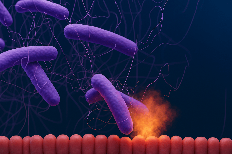
Serine proteases, in Leishmania species, help the parasite enter and grow in host macrophages. Additionally, they play a role in moving from the form of parasite that’s injected into humans through the bite of sandfly, to the form that lives in macrophages. This helps the parasite resist oxidative stress. Therefore, disruption of these serine proteases may help an infected person fight infection. To study this further, the researchers in this study explored how pretreatment with serine protease inhibitors affected the infection rate and survival of Leishmania amazonensis in host cells.
Testing the Inhibitors
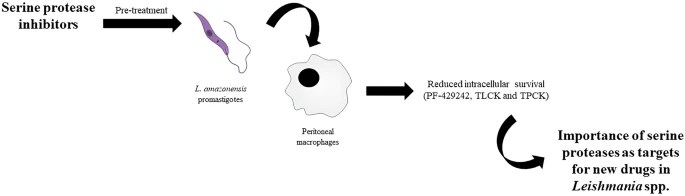
To conduct this experiment, researchers pretreated the human form of L. amazonensis with different concentrations of inhibitors (Figure 1). The known serine protease inhibitors used in this experiment were benzamidine, PF-429242, PMSF, TLCK and TPCK. Once pretreated, the cells were then used to infect peritoneal macrophages (immune cells found in the fluid between the abdominal wall and the organs of the abdomen) from mice. These macrophages were either pretreated or not pretreated with interferon gamma (IFN-γ), which enhances macrophages ability to respond to infection. After incubation, the number of parasites in each of the macrophages was measured.
Does Pretreatment Work?
What researchers discovered was that both benzamidine and PMSF pretreatment have no effect on entry of the parasite into the macrophage or on survival of the parasite within these host cells. This is as expected, however, as previous experiments demonstrated that benzamidine and PMSF have no effect on the parasites. In contrast, pretreatment with TLCK had no effect on parasite entry into the host cell, but did slightly reduce the survival of the parasite at 48 and 72 hours. Similarly, both PF-429242 and TPCK showed no effect on parasite entry, but did alter the survival of parasites throughout the first 72 hours.
Ultimately, inhibition of serine proteases can compromise survival of the parasite within the host cell, without altering the entry of these parasites into cells. These results demonstrate the potential use of serine protease inhibitors for treatment of Leishmaniasis.
Link to the original post: de Almeida Machado, P., Gomes, P.S., Coimbra, E.S. et al. Pretreatment with serine protease inhibitors impairs Leishmania amazonensis survival on macrophages. Parasites Vectors 18, 23 (2025). https://doi.org/10.1186/s13071-024-06630-w
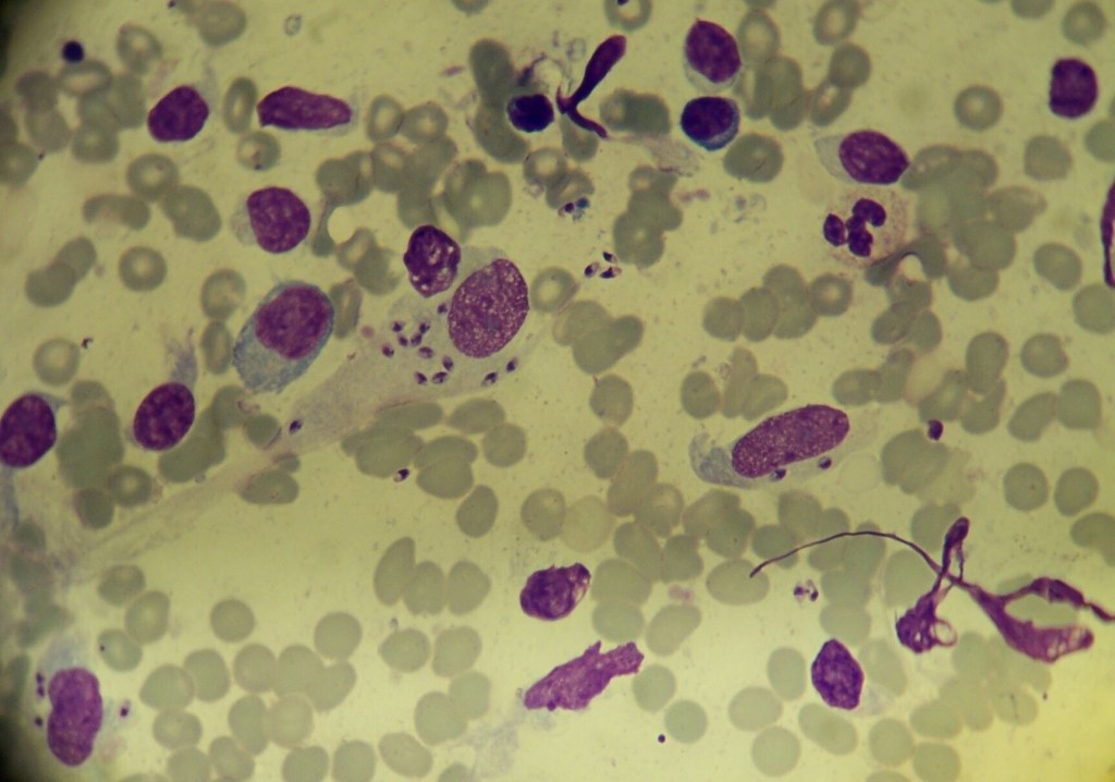
Featured image: Extracellular and intracellular leishmania parasites in a patient with cutaneous leishmaniasis | Source: Carcarrot, CC BY-SA 4.0 <https://creativecommons.org/licenses/by-sa/4.0>, via Wikimedia Commons