
Breaking down the microbiology world one bite at a time
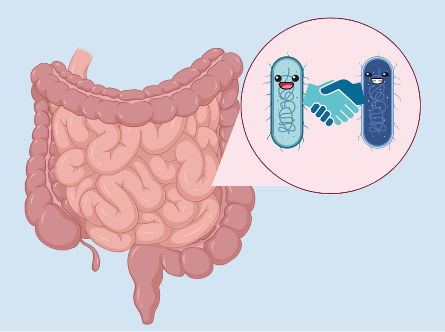
Healing in tandem
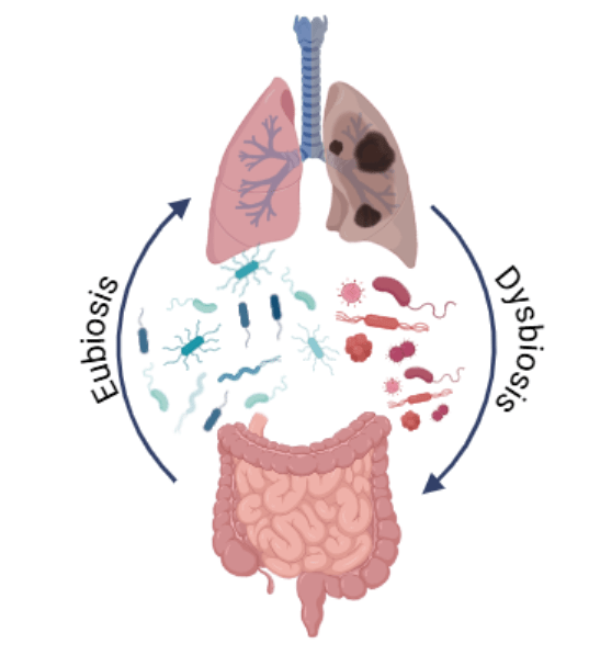
The gut microbiome consists of a vast and diverse community of microorganisms. The interaction among these microorganisms with the host’s tissue and immune system is a delicate balance to keep both the host and the microbes healthy and happy. When this balance is disrupted, such as through frequent antibiotic usage, diseases can occur, such as ulcerative colitis (UC).
UC is caused by abnormal inflammatory responses and can often lead to ulcer formation in the gut. Although UC is commonly considered to be caused by a disrupted immune system, recent studies suggest that the composition of the gut microbiome helps reduce the symptoms of UC. However, it is still not well understood what exact bacterial species are responsible for providing benefits to the preservation of the gut’s perfectly balanced environment.
The researchers of this study identified two different bacterial species— Akkermansia muciniphila and Parabacteroides distasonis— that synergistically protect against colitis. However, the way that these beneficial bacteria were discovered actually started with a failed experiment.
While performing mouse experiments to understand how the immune system responds during the course of UC infection, the authors noticed that some of their mice were not able to develop colitis, despite having long antibiotic treatment. This was extremely odd, since the authors used average wild type mice, with no modifications to their immune system.
That’s when the authors noticed that the set of mice that were resistant to UC came from a different animal facility. This was intriguing, as this suggested that something in their environment, such as food and water sources, might be the cause of their UC resistance.
The first test the authors did was to determine if these resistant mice had any differences in their gut epithelial layer stability or their gut immunity. Indeed, the resistant mice had upregulated E-cadherin and claudin genes, which promote the integrity of the gut epithelium. They also found that the resistant mice had higher interleukin-10 levels, which also helps to protect against UC.
The next step in uncovering the cause of this resistance to UC in these mice was to determine if the microbiome was involved. To do this, the authors performed a fecal microbiota transplant using feces from the resistant mice to treat the susceptible mice . Excitingly, when the susceptible mice were given this treatment, they had much higher resistance to UC, with only mild symptoms. This was a clear sign that the main protecting agent for the resistant mice was located in the microbiota.
To determine what microbes were involved, the authors then performed 16S rRNA gene analysis, which is often used to identify the genus and species of organisms in a biological sample. In the resistant mice feces, the authors noticed that the abundance of A. muciniphila and P. distasonis were higher than the sensitive mice.

Although A. muciniphila and P. distasonis were prevalent in the resistant mice’s microbiota, the authors still had to prove that these bacteria were the reason that the fecal microbiota transplant protected the sensitive mice from UC. To do this, the authors supplemented susceptible mice with either A. muciniphila, P. distasonis, or both. Interestingly, mice that were only supplemented with one of the bacteria were not able to resist UC. Only mice that were given both of the bacteria were able to resist UC. This implies that the bacteria are working together in some way to provide this protective effect.
Since supplementation with these bacteria protected against UC, the authors also wanted to determine if they could protect mice from relapsing UC. Mice that were given A. muciniphila or both bacteria were able to protect the mice from the second UC induction.
To investigate this further, the authors also analyzed the immune response of the mice in the presence of A. muciniphila. They found that this bacteria increases the number of type 3 innate lymphoid cells, which are involved in the innate immune response in mucous membranes, such as the lining of the gut. These gave the authors ample evidence that A. muciniphila is helping to prevent relapsing UC by contributing to the immune response.
Despite the surge in microbiome research in recent years, there is still a lot to be learned about this complex community. Exploratory studies such as this one are key to understanding the complexities of these systems. Although more information is needed about the mechanism of how these bacteria are providing protection, this is a great step forward in the fight against UC.
Link to the original post: Gaifem J, Mendes-Frias A, Wolter M, Steimle A, Garzón MJ, Ubeda C, Nobre C, González A, Pinho SS, Cunha C, Carvalho A, Castro AG, Desai MS, Rodrigues F, Silvestre R.2024.Akkermansia muciniphila and Parabacteroides distasonis synergistically protect from colitis by promoting ILC3 in the gut. mBio15:e00078-24. https://doi.org/10.1128/mbio.00078-24
Featured image: Created by Jana Gomez using BioRender