Breaking down the microbiology world one bite at a time
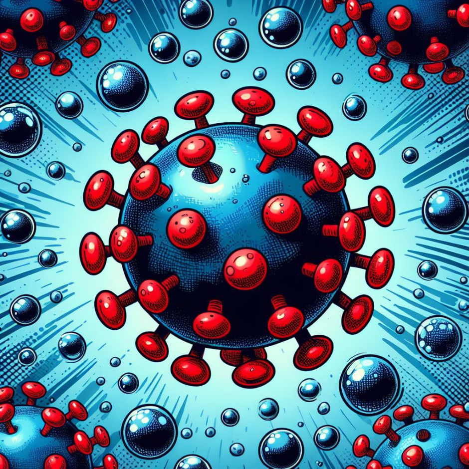
Viruses: the revolutionary power generators
Since ancient times, viruses have been known to cause many diseases. However, recent developments in science have facilitated the use of viruses for various biomedical applications, as viruses provide exceptional benefits for material design and synthesis. Firstly, due to their ability to encapsulate genetic blueprints within their protein coat, viruses can infect host cells and swiftly replicate a large number of identical copies of various materials. Secondly, viruses can be tailored to produce materials with peculiar biological functions and evolve into materials with novel capabilities. Thirdly, the ability to self-assemble provides the viruses with the advantage of forming a diverse range of supramolecular structures, which exhibit unique photonic and optical properties.
M13 BACTERIOPHAGE
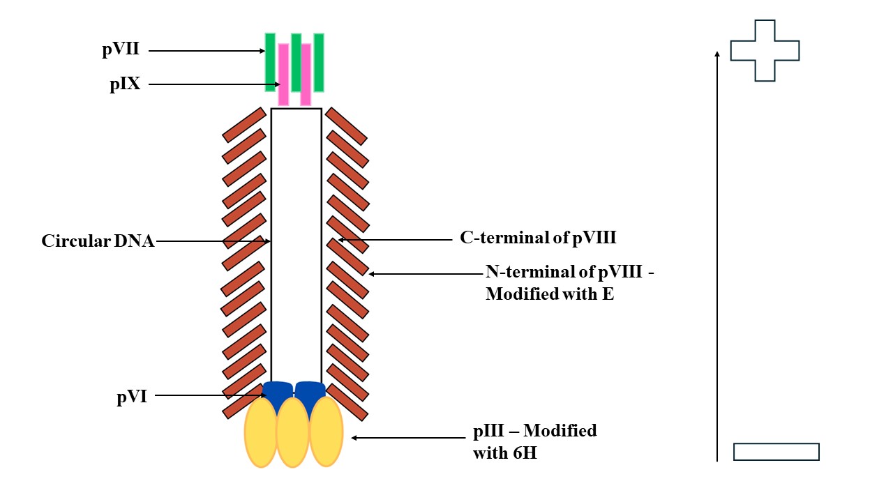
M13 phage is a member of the family filamentous bacteriophage (Ff phages), which infects bacteria having the fertility (F) factor. The virus particle has a diameter of 6 nm and is about 900 nm long. It has a circular single-stranded DNA that contains 6407 nucleotides. These nucleotides are encapsulated in the pVIII major coat protein and pIII, pVI, pVII, and pIX minor coat proteins. The pVIII protein has a positively charged C-terminal domain, an intermediate hydrophobic domain, and an N-terminal domain. The binding of the C-terminal domain with the genomic DNA of the phage creates a net negative charge on the surface of M13.
The M13 phage only infects bacterial species and is thus non-toxic to humans, making it a beneficial component in the fields of therapeutics, biosensing, immunoassays, and in vivo imaging. These phages can be easily engineered, for the development of various functional materials customized based on their functional needs. Further, its unique morphology and flexibility also enhance its performance. In addition to this, it also serves as a model virus particle for the phage display library. Many researchers have also used these phages as bio-templates for photocatalysts and dyes. Genetically engineered M13 phages have also been employed for the production of electricity in response to touch (triboelectricity) and pressure (piezoelectricity).
In the most recent work by Kim et al., major and minor coat proteins of the M13 phage were genetically tailored, inducing a unidirectional polarization in the phage particles. When subjected to heating, the a-helical structure of the phage proteins converted into random coils (in a reversible reaction), which consequently led to the generation of electricity. The electricity produced in response to a thermal (heat) stimulus is known as pyroelectricity. Pyroelectricity is associated with the change in the electric dipole moment of polar materials per unit volume under variations in temporal heat. Thus, a temporary voltage is generated by these materials when exposed to heating and cooling environments.
GENERATION OF PYROELECTRIC M13 BACTERIOPHAGES
The M13 phages cannot directly sense heat, however, thermal stimuli can induce structural changes in the phage particles. Therefore, the technique of genetic engineering was employed to modify the pIII and pVIII coat proteins of the phages to develop nano-sized, upright phage structures with unidirectional polarization. Along with the wild-type phages, these modified phages aligned vertically in a monolayer on a gold substrate.
The pIII minor coat protein, one of the most extensively used sites for polypeptide display, was modified with 6 molecules of the amino acid histidine (H). The pVIII major coat protein was altered by tuning the negative charge using varying numbers of the amino acid glutamate (E) on the protein surface, the structure-dependent pyroelectric potential of the phage could also be fine-tuned. As a result, heat (either from fire or from a laser) could denature the protein coating of the phage and generate electrical potential due to the unbalancing of the charges on the phage’s proteins. Although the maximum pyroelectric coefficient detected is very small (0.13 mC/m2/°C), the scientists intend to amplify the value by using the self-replicating property of the phage particles.
The authors further genetically modified the phage particles with various other molecules and investigated their potency as detectors of various volatile organic solvents, namely, methanol, ethanol, octanol, benzene, toluene, xylene, hexane, acetone, and diethyl ether. Thus, when modified with peptides tuned genetically to the specific solvents, the phage particles were able to generate pyroelectric responses depending on the concentration and type of chemical species.
This research has not only provided insights into the molecular mechanisms underlying the generation of electricity by various biomolecules, such as proteins, cells, and tissues, but will also pave the way for the development of novel biomaterials with applications in the fields of energy production, biosensors, medicine, pharmaceuticals, and environmental advancements.
!!Put caption of image also in alternative text!!
Link to the original post: H. Kim, K. Ontado, I. Chae, B. Lim, S. Ji, Y. Kwon, and S-W. Lee, Virus-based pyroelectricity, Advanced Materials, 35(46), e2305503, November 2023.
Additional sources:
1. R. Wang, H-D. Li, Y. Cao, Z-Y. Wang, T. Yang, and J-H. Wang, M13 phage: A versatile building block for a highly specific analysis platform, Analytical and Bioanalytical Chemistry, 415(18), 3927-3944, July 2023. DOI: 10.1007/s00216-023-04606-w.
2. D. Stopar, R.B. Spruijt, Cor J.A.M. Wolfs, and M.A. Hemminga, Protein-lipid interactions of bacteriophage M13 major coat protein, Biochimica et Biophysica Acta (BBA) – Biomembranes, 1611(1-2), 5-15, April 2003. DOI: 10.1016/S0005-2736(03)00047-6.
Featured image: Created by the author using Canva