Breaking down the microbiology world one bite at a time
Zika: The Brain’s Nemesis
In recent years, the Zika virus has made headlines around the world, largely due to its connection to serious birth defects like microcephaly, where babies are born with abnormally small heads and brain damage. A recent study explored the effects of Congenital Zika Syndrome (CZS) by looking at how these viruses interact with the cells in the brain. The researchers focused on how the Zika virus influences the formation of connections between brain cells, known as synapses, and the immune response in the brain, potentially leading to neurodevelopmental disorders such as Autism Spectrum Disorder (ASD).
The developing brain relies on a complex interplay of different types of cells, each playing a crucial role. To understand the results of this study, it is important to know a little about the brain cells involved. Neural progenitors are early-stage brain cells responsible for making new brain cells, particularly during brain development. Neurons are specialized cells in the brain and nervous system that transmit information through electrical and chemical signals. They communicate with each other through synapses, which are tiny bridges connecting them. Proper synapse formation is crucial for healthy brain function, affecting everything from learning to behaviour. Finally, astrocytes are a type of non-neuronal (glial) cells that support and protect neurons.
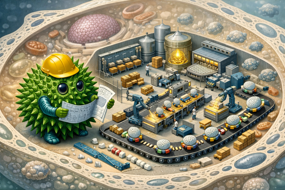
To understand the effects of the Zika virus on brain cells, the researchers took skin cells from children with CZS and transformed them into induced pluripotent stem cells (iPSCs), a special type of stem cell that can become almost any kind of cell in the body. The scientists then used a special process to differentiate these iPSCs into neural progenitor cells (NPCs), which are the building blocks for neurons and astrocytes.
The study revealed several key ways in which the Zika virus negatively impacts the formation of synapses in neurons. First, they found that the levels of important proteins, like presynaptic and postsynaptic proteins, were lower in neurons derived from children with CZS as compared to the healthy controls, thereby suggesting impaired synaptic function. Second, the study also revealed that the brain cells of CZS children had lower levels of important chemicals (neurotransmitters like glutamate) that help brain cells communicate. Astrocytes, which are the support cells for neurons, weren’t working as well in the CZS group. Specifically, some CZS patients had fewer astrocytes, and the astrocytes they had produced less glutamate and more inflammatory molecules. This is important because astrocytes help the neurons function properly. Finally, the CZS patients with fewer astrocytes were also the ones diagnosed with ASD, suggesting that problems with these support cells could be particularly harmful.

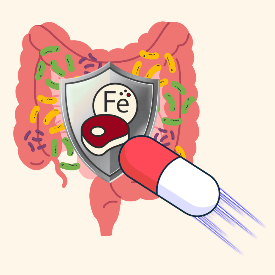
(Image Source: Made by the author using Canva)
A similar study by researchers at MIT and Harvard was done to understand the impacts of Zika and dengue viruses on brain cells. The study found that the Zika virus had a much more harmful effect on the brain cells than the dengue virus. The researchers created glial and neural progenitor cells in the lab from iPSCs and infected these cells with Zika and Dengue viruses to study how these viruses affect the brain cells.
They observed that the Zika virus readily infected NPCs and caused significant cell death, directly supporting the hypothesis that the Zika virus can disrupt the formation of neural cells by killing the cells responsible for generating new neurons. This is likely why the Zika virus leads to conditions like microcephaly in babies. Moreover, they found that glial cells did not die as readily as NPCs and instead could act as reservoirs for the virus within the developing brain, potentially contributing to ongoing viral activity and long-term consequences. Dengue virus, on the other hand, caused mild damage to brain cells, which wasn’t as severe as the damage caused by Zika.

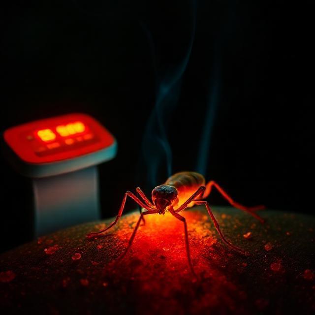
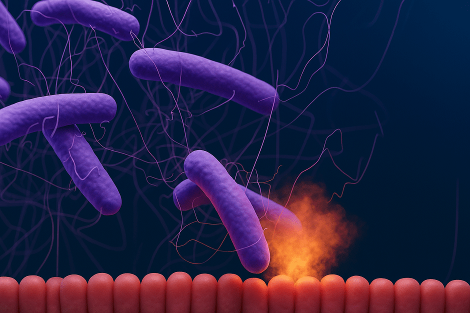
Mosquitoes are carriers of the Zika virus. When a mosquito bites the mother, the Zika virus enters her blood. It infects certain immune cells that carry it across the placenta, via the mother’s bloodstream, to the baby’s brain. The virus infects cells in the placenta, allowing it to bypass this protective barrier and infect the developing fetus’s brain. Fetal microglia, immune cells in the developing brain, are the ultimate target. (Image source: Created by the author using Canva)
These studies open the door to new research into how various harmful viruses, like the Zika and dengue viruses, interact with the brain. With this knowledge, researchers hope to develop more effective therapies to protect pregnant women and babies from the toxic effects of these viruses and better understand how they affect the brain in general. It is a step forward in understanding how viruses can affect human health, especially in the most vulnerable populations. The hope is that by targeting the specific responses of brain cells to these viruses, doctors and scientists can find ways to prevent brain damage, treat infected individuals, and even develop vaccines or therapies that prevent the viruses from causing such severe health problems.
Moreover, understanding how the Zika virus affects brain cells will help researchers identify potential long-term impacts on children born with CZS, including risks for developmental disorders like ADHD. This knowledge is crucial for developing strategies to support affected children and improve their outcomes. By learning more about these diseases, scientists are paving the way for better prevention, treatment, and protection against these dangerous viruses.
A question for the readers: Did you notice how both studies have used different approaches to understand the interaction of viruses with brain cells? Scientists in the first study tested the iPSCs from the skin cells of children with CZS. On the other hand, the researchers in the second study infected normal iPSCs with the viruses.
Additional source: J. Muffat, Y. Li, A. Omer, A. Durbin, I. Bosch, G. Bakiasi, E. Richards, A. Meyer, L. Gehrke, R. Jaenisch, Human induced pluripotent stem cell-derived glial cells and neural progenitors display divergent responses to Zika and dengue infections, Proc. Natl. Acad. Sci. U.S.A., 2018 June 18, 115(27), 7117-7122. DOI: 10.1073/pnas.1719266115.
Featured image: (Image source: Created by the author using Bing Image Creator)